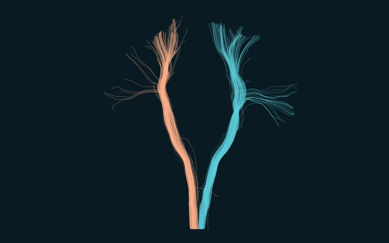
 DIFFUSION MRI TRACTOGRAPHYMRI
Corticospinal Tract Pathways
Follow diffusion MRI corticospinal pathways, then explore illustrative pyramidal and cervical crossings, anterior horn targets and lesions above the decussation.
Enter the labDICOM & 3D ANATOMY
Real image stacks, coronary cine, and interactive teaching models. Choose a region to open its full imaging workspace.
10 anatomy labs
DIFFUSION MRI TRACTOGRAPHYMRI Follow diffusion MRI corticospinal pathways, then explore illustrative pyramidal and cervical crossings, anterior horn targets and lesions above the decussation.
Enter the lab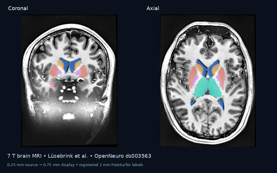 7T BRAIN MRI + REGISTERED ANATOMYMRI
Explore an ultrahigh-resolution brain MRI with linked slices, deep nuclei, atlas-based brainstem parts, ventricular opening landmarks and labeled cranial-nerve teaching guides.
Enter the lab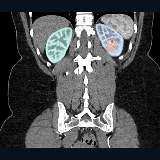 SOURCE CT + KIDNEY / TUMOR MASKSCT
Explore a thin-slice arterial CT volume with source kidney and tumor masks, provisional renal vessels and surrounding anatomy, linked CT views, and movable labels.
Enter the lab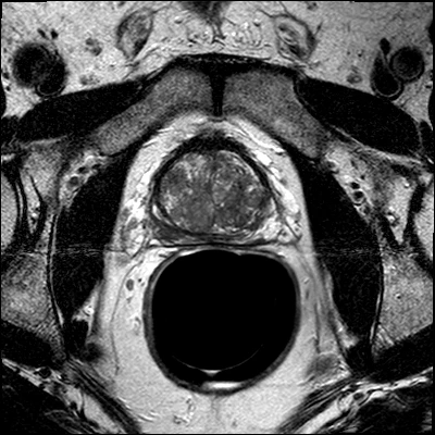 SOURCE T2 MRI · CASE 0006MRI
Explore the prostate and adjacent anatomy using linked MRI views and 3D models. Consider what the findings might represent, form your interpretation, then compare it with the teaching reference.
Enter the lab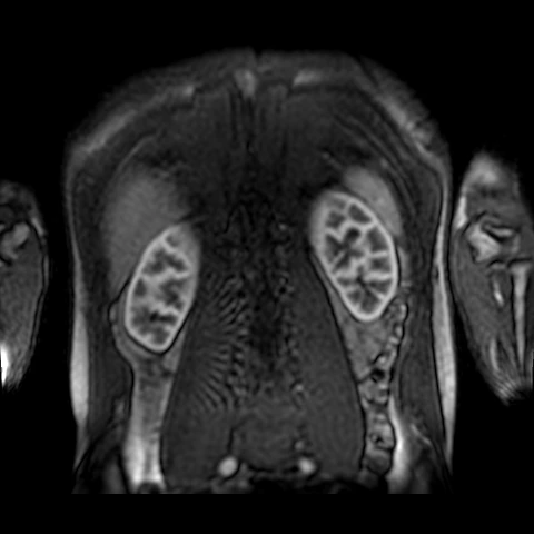 SOURCE RENAL DCE MRIMRI
Explore renal contrast enhancement through 160 time points, labeled native images, and an explicitly partial three-dimensional kidney model.
Enter the lab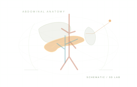 SCHEMATIC ILLUSTRATIONCT
Follow the pancreas through an image stack, then connect its anatomy with vessels and the biliary tree.
Enter the lab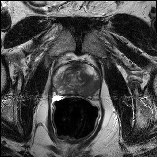 SOURCE MRIMRI
Connect T2-weighted MRI with the prostate, seminal vesicles, urethral segments, and pelvic vessels.
Enter the lab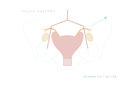 SCHEMATIC ILLUSTRATIONMRI
Explore linked pelvic MRI planes with reproductive anatomy and a named pelvic arterial branch map.
Enter the lab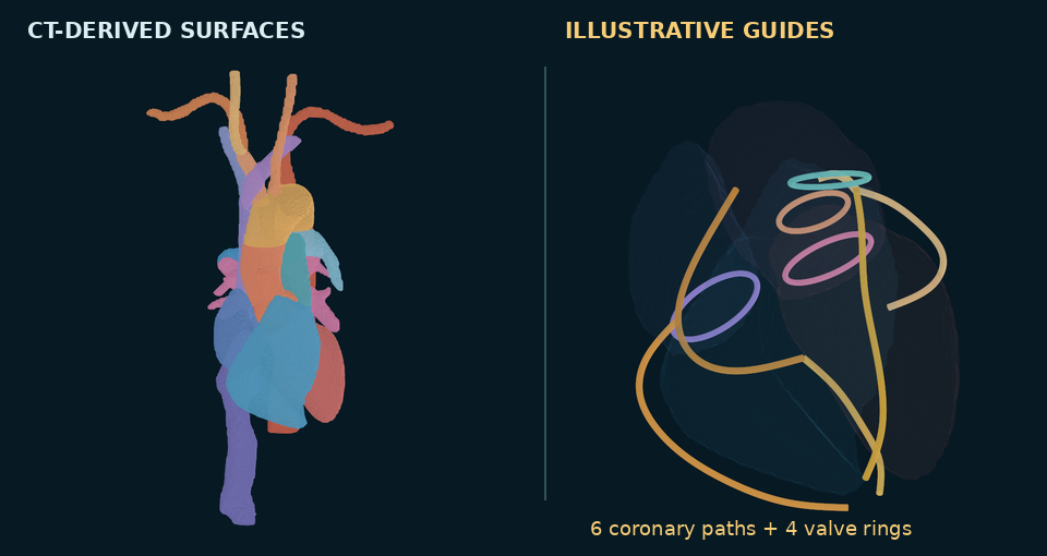 CT-DERIVED & ILLUSTRATIVE MODELSCT
Connect linked thoracic CT views with three-dimensional cardiac and vascular anatomy, saved labels, and guided exploration.
Enter the lab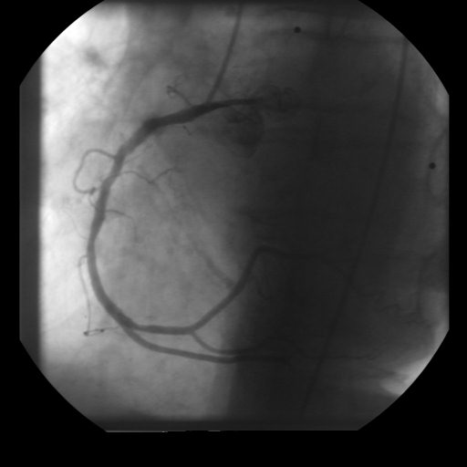 SOURCE ANGIOGRAPHYAngiography
Review coronary cine imaging frame by frame with labeled right coronary branches and a linked projection model.
Enter the labTry another structure or select all modalities and regions.
Learn the imaging planes and orientation labels before entering the viewer.