MRI / Male pelvis
MRI prostate with document interpretation
Explore the prostate and adjacent anatomy using linked MRI views and 3D models. Consider what the findings might represent, form your interpretation, then compare it with the teaching reference.
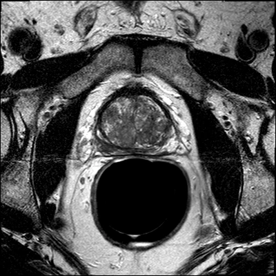 SOURCE T2 MRI · CASE 0006TCIA MRI + 3D source masks
GUIDED EXPLORATION
A route through the lab.
- 01
Browse 32 native axial T2 slices with linked coronal and sagittal oblique reformats. Review the same-patient DCE companion: 256 images across eight time points, with separate temporal and spatial controls and coordinate-linked crosshairs. Existing window/level, pan, zoom, cine and layout controls are retained. Full-grid 3D surfaces and up-to-8K exports do not increase acquired MRI resolution.
- 02
Explore 12 anatomy layers with slice-linked labels. Read the provenance for each layer: additional atlas identities are inferred, and small structures may not be independently resolved at 3 mm slice thickness.
- 03
Write or dictate your scan interpretation. Submit a local snapshot, then compare it side by side with a standardized, source-based teaching note and a reporting-element self-review. No automated clinical accuracy score is assigned.
- 04
Create tags, calibrated lengths, angles, signal ROIs and draft contours. Save or export your annotations, interpretation, comparison, PNG images and full-detail research STL surfaces.