INSTRUCTIONAL POSTER · PROJECT OVERVIEW
Generative Artificial Intelligence–Assisted Development of an Interactive Three-Dimensional Cardiovascular Anatomy Explorer
This instructional poster presents the development of a browser-based cardiovascular anatomy explorer that integrates computed tomography images with interactive three-dimensional reconstructions for medical and physician assistant education. The platform is designed to help learners connect cross-sectional imaging with the spatial relationships of cardiovascular structures.
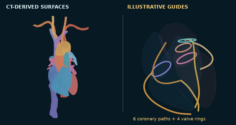
Generative artificial intelligence supported application development, while automated segmentation generated preliminary anatomical models from a publicly available contrast-enhanced thoracic computed tomography dataset obtained from The Cancer Imaging Archive. Linked axial, coronal, and sagittal views allow simultaneous exploration of cardiac chambers, major arteries and veins, the esophagus, and vertebral levels. Learners can rotate models, adjust structure visibility and opacity, and display imaging planes within the reconstruction.
Teaching features include movable labels, measurement tools, image bookmarks, and saved annotations. Faculty-created tags appear within the anatomy panel, with editing and deletion restricted to the owner. Coronary artery courses and heart valve positions are explicitly identified as illustrative guides where the scan does not support reliable segmentation.
The presentation demonstrates how these features can support guided anatomical exploration, image–structure correlation, and faculty-directed review. It also addresses the distinction between image-derived anatomy and illustrative models, emphasizing the need for expert review of automated segmentations. This project represents an educational development initiative; formal assessment of anatomical accuracy, usability, and effects on learner performance remains necessary.