CARDIOVASCULAR CT · DEVELOPMENT & LEARNING

Connect the image.
Explore the anatomy.

Cardiovascular anatomy and viewer skills for first-year and clinical-year medical students, using linked CT views and an interactive three-dimensional teaching model.

The explorer opens within ImagingNest. Proposed learning activities and preliminary models require educator review.

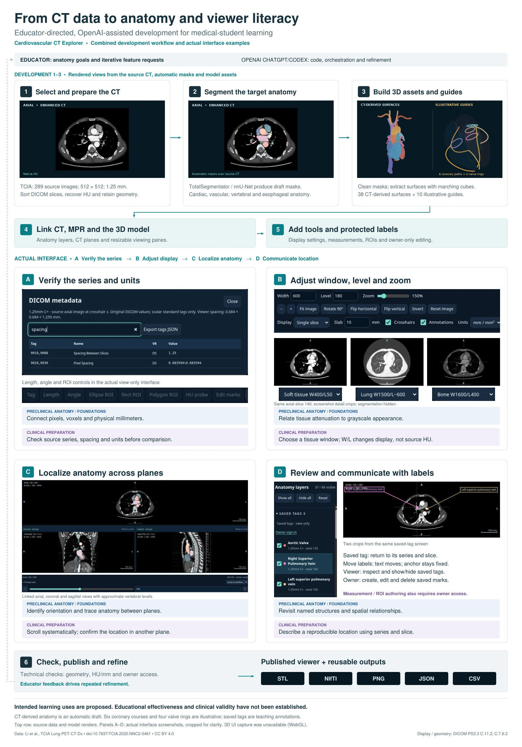
Workflow poster connecting CT preparation, segmentation, 3D reconstruction, linked views, viewer tools, and technical checks. From CT data to anatomy and viewer literacy Open the PDF
Selected CT series
289 images
Slice thickness
1.25 mm
CT-derived surfaces
38 draft models
Illustrative guides
10 coronary & valve

HOW IT WAS DEVELOPED

Educator goals.
Iterative development.

Educators defined the anatomical emphasis and requested features. OpenAI ChatGPT and Codex supported application coding, workflow orchestration, interface refinement, and troubleshooting through iterative natural-language instructions.

  1. Select and prepare the CT

    Select publicly available, de-identified TCIA data. Extract acquisition metadata, assess vascular enhancement and slice continuity, sort DICOM images, recover Hounsfield units, and retain spatial geometry.

  2. Segment the target anatomy

    Use TotalSegmentator / nnU-Net to generate draft masks for cardiac, vascular, vertebral, and esophageal anatomy. Automated boundaries remain preliminary.

  3. Build surfaces and guides

    Clean masks and extract three-dimensional surfaces with marching cubes. Keep CT-derived surfaces distinct from the six illustrative coronary courses and four illustrative valve guides.

  4. Link images and anatomy

    Integrate the model with linked axial, coronal, and sagittal views, selectable anatomy layers, adjustable opacity, slice planes, and resizable viewing panes.

  5. Add tools and protected labels

    Add display settings, saved tags, bookmarks, measurements, and region-of-interest tools. Owner access controls annotation and measurement authoring; learners can inspect saved teaching marks.

  6. Check, publish, and refine

    Check spatial alignment, attenuation and physical units, exported-file integrity, and owner permissions. Educator feedback informs refinement. Reusable outputs include STL, NIfTI, PNG, JSON, and CSV.

QUANTITATIVE SOURCE EXTRACTION

Choose the source.
Document the trade-offs.

Image counts, slice thickness, and slice positions informed source selection. Enhancement was assessed qualitatively. These findings describe source characteristics and development outputs.

CT series assessed during development
Source characteristicInitially selected seriesSelected companion series
Image count600289
Slice thickness0.625 mm1.25 mm
Vascular enhancementLimitedContrast-enhanced
Missing slice positions1 detected0 detected
Reconstruction decisionReplaced after source reviewSelected for reconstruction

The selected series has a 512 × 512 image matrix. Slice thickness and in-plane pixel spacing are separate quantities; check source metadata before interpreting physical measurements.

From initial segmentation to the expanded model

Initial segmentation generated 20 named anatomical surfaces. The expanded workflow documents 38 CT-derived surfaces, with additions including the esophagus and vertebral references spanning C7–L3. Six coronary artery course guides and four heart valve guides are explicitly illustrative because reliable segmentation was unavailable.

FIRST-YEAR & CLINICAL-YEAR MEDICAL STUDENTS

One workspace.
Different learning goals.

Educators can tailor anatomical emphasis, saved annotations, and review tasks. Learners control image navigation and model exploration. The activities below are proposed uses, with progression directed by an educator.

Four ways to work with the explorer
Viewer taskFirst-year focusClinical-year focus
Verify the series and unitsConnect pixels, voxels, and physical dimensions in millimeters.Check source series, spacing, and units before comparing views or measurements.
Adjust window, level, and zoomRelate tissue attenuation to grayscale appearance.Choose a tissue window and explain why display adjustments do not change source Hounsfield units.
Localize anatomy across planesIdentify orientation, name structures, and trace spatial relationships.Scroll systematically and confirm an anatomical location in a second plane.
Review and communicate with labelsRevisit named structures and compare their relationships.Describe a reproducible location using the series, slice, and imaging plane during guided discussion.

Individualization through educator control

OpenAI-assisted development offers a way to translate teaching goals into tailored radiology resources. Educators choose content and review tasks; learners choose the pace and views they explore. Automated adaptation based on learner performance has not been demonstrated.

Teaching annotations with a stable reference

Saved tags return to their series and slice. Moving label text preserves its anatomical anchor. Viewers can inspect and show or hide saved marks; creating, editing, or deleting marks and authoring measurements or regions of interest require owner access.

THE COMPLETE WORKFLOW

From CT data to
viewer literacy.

The combined figure pairs the development sequence with source-data renders and actual interface examples. Open the PDF to examine the details at full resolution.

Six-stage development workflow and four viewer activities: verify series and units; adjust window, level, and zoom; localize anatomy across planes; review and communicate using saved labels. Equivalent descriptions appear in the sections above.
Top row: source CT, automatic masks, and model renders. Panels A–D: actual interface screenshots cropped for clarity. The figure distinguishes CT-derived draft anatomy from illustrative guides.

DATA, PRIVACY & EVALUATION

Keep the source
and scope visible.

Public teaching data

The prototype uses publicly available, de-identified CT images from The Cancer Imaging Archive. Only de-identified teaching data are appropriate for this workflow. Keep patient identifiers out of annotations, screenshots, exports, and AI prompts.

Use of public data does not by itself establish HIPAA compliance of an entire application or workflow. A compliance claim requires verification of the actual data handling and safeguards.

HHS guidance on HIPAA de-identification

What the prototype demonstrates

The completed prototype demonstrates a development approach and technical integration. Automated segmentations remain preliminary. Anatomical accuracy, usability, clinical validity, and educational effectiveness require formal evaluation.

Technical checks and counts of images or surfaces do not establish learning gains. Faculty remain responsible for reviewing anatomical content and making instructional decisions.
Source and standards

Start with the source image.

Open the explorer and choose a learning activity above.

Open the cardiovascular explorer

Find your next connection.

Search the pages in ImagingNest AI.