CT / Abdomen
Renal CT & surgical anatomy
Explore a thin-slice arterial CT volume with source kidney and tumor masks, provisional renal vessels and surrounding anatomy, linked CT views, and movable labels.
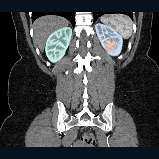 SOURCE CT + KIDNEY / TUMOR MASKSNative CT + source kidney & tumor masks
GUIDED EXPLORATION
A route through the lab.
- 01
Locate the source tumor mask in the axial CT. Follow its extent through adjacent slices, then compare its coronal and sagittal appearances.
- 02
Rotate the kidney and tumor models. Lower the affected kidney opacity to inspect the tumor relationship to its parenchyma. Compare the provisional renal vessels, proximal SMA, adrenal glands and upper ovarian veins with the source CT. Use Anterior bowel to inspect small-bowel loops and the duodenum in relation to the kidneys. Use Vessel coverage to identify unresolved adrenal vessels and ovarian arteries. The Partial nephrectomy measurement workspace lets you inspect provisional mask gaps, place CT landmarks and export a review worksheet. These measurements are not safe resection margins.
- 03
Adjust window and level, zoom, pan, or inversion. Use length, angle and HU ROI tools; add personal tags and bookmarks.
- 04
Drag labels and personal tags, or resize the 3D and CT panels. Export labeled images, model surfaces or personal annotations with the dataset attribution.