MRI / Male pelvis
Prostate & male pelvis
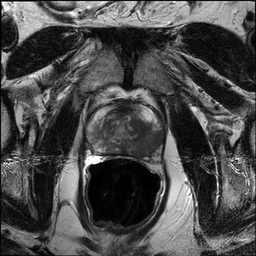
Connect T2-weighted MRI with the prostate, seminal vesicles, urethral segments, and pelvic vessels.
 SOURCE MRIMRI · DICOM + 3D
This case was acquired with an endorectal coil, which distends the rectal lumen. Organ envelopes are draft outlines; ducts, ureters, urethra, and vessels are schematic. Some paths extend outside the scan.
GUIDED EXPLORATION
A route through the lab.
- 01
Browse the acquired axial, coronal, and sagittal T2 series. Observe how one structure appears in each plane.
- 02
Select a draft organ outline and use Locate on MRI to compare the boundary with the source images.
- 03
Choose the Urethra, Reproductive, or Ureters preset. Read the structure description before interpreting its position.
- 04
Open the Vessels preset. Select a named artery or vein to follow its displayed origin or drainage route.