MRI / Abdomen
Contrast-enhanced renal MRI
Explore renal contrast enhancement through 160 time points, labeled native images, and an explicitly partial three-dimensional kidney model.
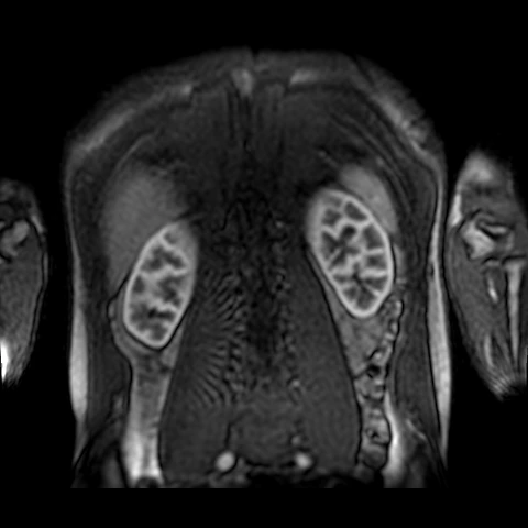 SOURCE RENAL DCE MRIDCE MRI + partial 3D anatomy
Source: Peter Thelwall, Newcastle University, Renal DCE scan data - anonymised (2020), version 2, CC BY 4.0. Vol B includes three 8-mm coronal-oblique slices plus one axial-oblique slice. Manual renal surfaces are partial and unvalidated; this is not a complete excretory MR urogram.
GUIDED EXPLORATION
A route through the lab.
- 01
Compare the three acquired coronal-oblique images and the separate axial-oblique image. These are four spatial positions repeated over time, not 640 anatomical slices.
- 02
Select a kidney or landmark to locate it at reference time point 60. Rotate the partial model and inspect its open boundaries.
- 03
Play the contrast-enhancement cine. Adjust window and level, zoom, and pan; compare signal changes using the fixed sample ROI curves.
- 04
Create a personal draft tag, calibrated length, angle or rectangular ROI. Save in your browser or export JSON, CSV, and labeled PNG images.