CT / Abdomen
Pancreas & abdominal vessels
Follow the pancreas through an image stack, then connect its anatomy with vessels and the biliary tree.
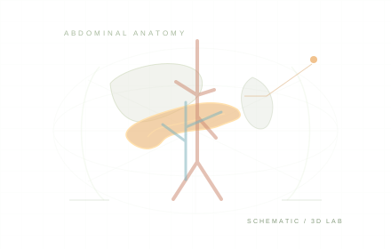 SCHEMATIC ILLUSTRATIONCT · DICOM + 3D
Use the viewer’s model notes to distinguish reconstructed structures from schematic anatomy. A teaching model does not establish a clinical diagnosis.
GUIDED EXPLORATION
A route through the lab.
- 01
Start with the CT stack. Scroll slowly and locate the pancreas over several consecutive slices.
- 02
Turn on the pancreas in the 3D view. Rotate the model to understand its position within the abdomen.
- 03
Add arterial, venous, and biliary structures one group at a time. Read the provenance notes for each teaching model.
- 04
Tag an anatomical relationship in the image and in 3D. Compare the two views before moving on.