MRI / Female pelvis
Uterus & female pelvis
Explore linked pelvic MRI planes with reproductive anatomy and a named pelvic arterial branch map.
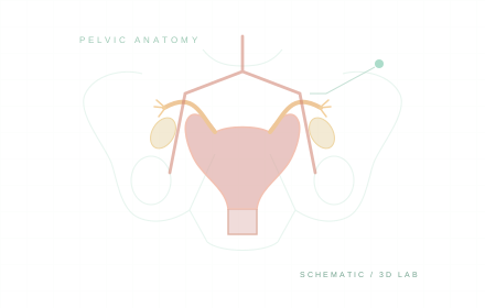 SCHEMATIC ILLUSTRATIONMRI · DICOM + 3D
Small reproductive structures and vascular branches may be schematic teaching aids rather than visible, segmented structures in the MRI. Read the viewer’s source and coverage notes.
GUIDED EXPLORATION
A route through the lab.
- 01
Orient yourself in the linked MRI planes. Establish anterior, posterior, superior, and inferior relationships.
- 02
Add the pelvic teaching models and rotate to an anterior or lateral view.
- 03
Toggle the uterine tubes to review their illustrated parts. Check whether the viewer identifies a structure as schematic.
- 04
Select the internal iliac arteries and named branches. Use the vessel names and routes to identify each branch.